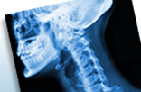
70 year old female patient with a large, palpable swelling to the left side of the neck.
CT Scan of the neck with contrast agent was done on a 40 slice Sensation CT scanner with a slice thickness of 5.5 mm and reconconstructed with a 2.5 mm thickness.
There is a 5.2 x 4.1 x 5.8 cm well-defined intensely enhancing soft tissue density mass noted in the left carotid space at the level of the left carotid artery bifurcation.
There are non-enhancing areas noted within the mass, suggestive of necrosis. This mass displaces the left internal carotid artery and left jugular vein laterally.
Overall features are suggestive of left carotid body tumor.
(Courtesy Southern Medical Clinic Ltd/San Fernando).
Other Cases
2009